baby chest x ray technique
A chest radiograph for a 12-year-old female is an embarrassing ordeal. They can also help confirm that medical.

Approach To Pediatric Chest X Rays Youtube
But newborns unlike older infants and children have.

. While doing the X-ray Asepsis precautions. The neonatal chest X-ray R. Most neonatal chest X-rays are AP films unless the baby is made to lie prone Lucency of soft tissue shadow - darker the soft tissue more.
The anteroposterior AP diameter of the neonatal chest is almost as great as its transverse diameter giving the chest a cylindrical configuration. X-ray exams are used to help diagnose a wide variety of injuries and illnesses in children. A chest X-ray can help doctors find the cause of a cough shortness of breath or chest pain.
If exam is the initial chest image please include the following in tech comments. X-rays are used throughout the body. Whilst many of the radiological appearances are relatively non-specific integration of the clinical features with the X-ray.
Birth Weight Gestational Age and Vaginal or Cesarean. In contrast most 12-year-old males have little modesty about their chests. Pediatric Chest Screen 70-80 DIGITAL OPTIMUM kVp Universal CR Technique Chart using a standard 21 LgM Part View kV mAs kV mAs kV mAs Abdomen AP Grid 85 10 -15 85 20 - 25 85 30 - 40 Ankle AP 70 18 70 2 70 25 Ankle Obl 70 16 70 18 70 22 Ankle Lat 70 15 70 16 70 2 Chest -Adult AP 400 - tt -72 85 2 - 25 85 32 - 4 90 5 - 64.
The degree of ionizing radiation is not considered dangerous to human health so X-rays can be considered a good alternative to ultrasound 1 computed. 1 the relative size of a lung or hemithorax 2 the degree of radiolucency of the lung and 3 the pulmonary vascularity or blood flow to the lung. No evidence of pneumonia.
Chest lateral 180 cm. 1A Chest radiographs of two different patients. Chest X-rays can show a swallowed foreign object such as a coin.
When focused on the chest it can help spot abnormalities or. When X-rays of the ribs the state of the bone mechanism is visualized and the spine can be partially seen. It is often the first type of imaging used to identify sources of pain evaluate traumatic injuries and locate a foreign body.
The technique factors used should be chosen based on the clinical indication patient size and anatomical area scanned and the equipment should be. In this study the exposure technique of 65 kVp and 16 mAs was chosen as a reference image due to this technique being near the suitable exposure uses in. This technique represents the expansion in two dimensions only.
Initial chest radiograph obtained immediately after surgery A shows normal contour of superior mediastinumFollow-up chest radiograph obtained. PA or AP and left lateral supine in infants upright in children or when requested. Frequently for a single child both radiographs are requested simultaneously.
Chest x-ray is the most commonly used imaging exam for evaluating the chest. 37 years average 5 years. As newborn chest radiographs are taken in the AP plane the normal cardiothoracic ratio.
The chest X-ray is the most frequently ordered radiological. Lie on an X-ray table on your side with your ear glued to. The degree of rotation is best assessed by comparing the length of the anterior ribs visible on both sides.
A 9-month-old girl with history of atrioventricular septal defect who underwent repair and heart block requiring permanent pacemaker now receiving warfarin. However all children are modest to some degree about having their genitals or backsides exposed after ages 4 to 5. Arthur X-ray and Ultrasound Department Leeds Infirmary Leeds UK Summary The chest X-ray is the most valuable imaging modality in the assessment of the neonate with respiratory distress.
Chest lateral supine 110 cm. X-ray Imaging for Pediatrics. Plain X-ray shows the existing damage to the internal organs and the whole chest.
7725a a Use automatic exposure control 500 speed for chestabdomen else 400 speed at specified kVp when practical. Prevent hypothermia. 812 years average 10 years.
It can detect signs of pneumonia a collapsed lung heart problems such as an enlarged heart and broken ribs or lung damage after an injury. Radiographs of the chest and the abdomen are the most commonly requested diagnostic X-ray examinations undertaken in neonatal intensive care units. AP and lateral chest x-ray demonstrates minimal peribronchial cuffing likely related to IV fluids.
The age groups were based on exposures suitable for tissue thickness in the direction of the X-ray beam of a patient of averagestandard size in that age group for each projection. These images can be obtained either as two separate exposures one of the chest and one of the abdomen or as a single exposure to. Chest Routine chest.
1317 years average 15 years. An X-ray is an imaging test that uses small amounts of radiation to produce pictures of the organs tissues and bones of the body. 9025a a Use automatic exposure control 500 speed for chestabdomen else 400 speed at specified kVp when practical.
Quieten the baby to avoid swings in respiratory depth While reading the X-ray Read schematically Do not jump to the diagnosis - you will miss important additional findings Make differential diagnosis and correlate clinically Write age in hoursdays on the X-ray. Pulmonary aeration abnormalities are best evaluated on the chest radiograph by observing the following criteria. AP supine and lateral cross-table o Newborn Initial Chest.

Abdomen X Ray Radiographie

Neonate Chest Supine View Radiology Reference Article Radiopaedia Org

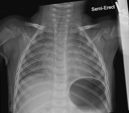
Pleural Effusion Undergraduate Diagnostic Imaging Fundamentals

Pediatric Chest Horizontal Beam Lateral View Radiology Reference Article Radiopaedia Org

Chest Radiograph Pediatric Radiology Reference Article Radiopaedia Org

Pin On Radiology Views Pathology And Such

Pin On Ir Tech

Pediatric Chest Supine View Radiology Reference Article Radiopaedia Org
2

Neonatal Radiography Part 1 Nomal Findings And The Basics Youtube
A Pediatric Chest X Ray With The Pb Shield The Circles Are The Download Scientific Diagram

Pin By Eve Emmanuelle Roy Hebert On T I M Diagnostic Imaging Med Student Medical Field

The Forbidden Chest X Ray Tension Pyopneumothorax The American Journal Of Emergency Medicine

Chest X Ray Of A 6 Month Old Child With An Icd The Active Can Is Download Scientific Diagram

X Ray Imaging For Covid 19 Patients
2
2
Underinspiration And Poor Positioning Mimicking Lung Pathology In A Pediatric Patient Radiology Case Radiopaedia Org
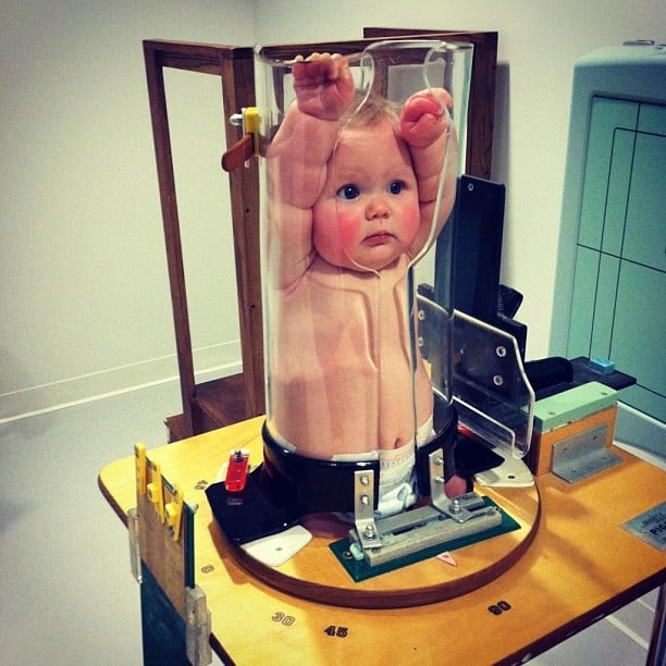
Ce4rt Guide For X Ray Techs To Immobilize Pediatrict Patients